


Sjogren's syndrome
Sjogren’s syndrome (SS) is a chronic autoimmune disease which affects the salivary and lacrimal glands, as well as other exocrine glands, leading to functional exhaustion and dryness of mucous membranes, and is often associated with systemic extraglandular manifestations. SS can be primary or secondary to other autoimmune diseases (SLE, rheumatoid arthritis, scleroderma, biliary cirrhosis, etc.). In the primary syndrome, which is very rare in children, the sicca component is the dominant manifestation and is associated with lymphocytic infiltration of the lacrimal and salivary glands. In 50% of patients, connective tissue disease manifestations dominate.
6.2.1 EPIDEMIOLOGY
Frequency: SS is the most common, after RA, autoimmune disease. In the U.S., about 500.0000-2.000.000 people suffer from SS. In the rest of the world, the prevalence of SS is similar to the U.S.. In children, primary SS is rare. Only a few cases have been reported(Chudwin DS et al, 1981; Kraus A and Alarcon-Segovia D, 1988).
Age: In the U.S., SS mainly affects women in the fourth and fifth decade of life and generally people of any age, but is relatively rare in children. The youngest patient with SS reported is a girl aged 3 years.
Gender: The ratio of female / male is approximately 9:1.
6.2.2 AETIOLOGY
Multiple causative factors are likely to be involved in the pathogenesis of SS.
Sex hormones: As in other autoimmune diseases, SS is prevalent in women.
Genetic factors: Patients with SS and HLA-B8, Dw3 DR3 exhibit a high frequency of autoantibody production and extraglandular manifestations. Most of them, also carry the DQA1501 allele. Family members of patients with SS have a higher frequency of SS.
Infections:During primary infection, the Epstein-Barr virus (EBV) reproduces in the salivary glands, where it remains dormant. EBV-DNA has been isolated in the salivary glands and saliva of patients infected with EBV.
Autoantibodies: Anti-Ro (SS-A) and anti-La (SS-B) are associated with earlier onset, longer duration and more extraglandular manifestations of primary SS.
Immunological factors: IL-1, IL-6 and TNF are produced in the salivary glands in the infiltrating lymphocytes, and epithelial cells, in situ. Their presence is an indication that the infiltrating lymphocytes (e.g. mainly CD4 T cells) play a role in perpetuating the immunological process of SS.
CONDITIONS ASSOCIATED WITH SJOGREN’S SYNDROME
- Vasculitis
- Dryness syndrome
- Bilateral parotid gland enlargement
- Achlorhydria
- Hepatomegaly
- Hashimoto’s thyroiditis
- Celiac disease
- Lymphoid myositis
- Pancreatitis
- Hyposthenuria and renal tubular acidosis
- Connective tissue diseases
- Juvenile idiopathic arthritis (Franklin DJ et al, 1986)
- Polyarteritis nodosa
- Polymyositis
- Systemic lupus erythematosus (Franklin DJ et al, 1986)
- Scleroderma
- Hypergammaglobulinemia purpura (Celada A et al, 1980)
- Raynaud’s phenomenon
- Chronic active hepatitis
- Autoimmune hepatitis (Bartunkova J et al, 1999)
- Primary biliary cirrhosis
- Malignant diseases
- Pseudolymphoma
- Lymphoma
LABORATORY FINDINGS ASSOCIATED WITH SJOGREN’S SYNDROME
- Autoantibodies
- Rheumatoid factors
- ANA
- Lupus cells
- Antibodies to SS-A, SS-B and RAP
- Antibodies to salivary ducts
- Anti-tissue antibodies
- Macroglobulinemia
6.2.3 CLINICAL MANIFESTATIONS
SS is characterized by the following triad of signs and symptoms:
- Sicca syndrome, manifested mainly by keratoconjunctivitis sicca (dry eyes) and xerostomia
- A connective tissue disease(usually SLE)
- Autoantibodies in high titres, usually ANA (anti-Ro/La), or RF.
In children, the first manifestation of primary SS is usually recurrent enlargement of the parotid glands (Siamopoulou-Mavridou A et al, 1989; Anaya JM et al, 1995; Drosos AA et al, 1997) or enlargement of the salivary glands.
6.2.3.1 EXOCRINE GLAND INVOLVEMENT
The main manifestations of exocrine gland involvement are:
- Keratoconjunctivitis sicca(xerophthalmia)
- Xerostomia
- Salivary gland enlargement.
Exocrine glands of the upper respiratory tract can also be affected. The involvement of the exocrine glands of the skin account for dry skin.
6.2.3.1.1 KERATOCONJUNCTIVITIS SICCA
It usually develops silently over several years and may be complicated by corneal ulceration and blepharitis. It is characterized by dryness and a foreign body sensation in the eyes. These symptoms are often exacerbated in the afternoon. Patients often report that they have "sawdust, sand or gravel," rather than dryness in the eyes, but curiously, when crying, they shed tears. This is due to a reduction of the aqueous component of tears and is one of the most common causes of dry eyes, especially in the elderly (Schein OD et al, 1999). The risk of developing sicca symptoms may depend on estrogen, since primary SS is more common in women (96%) (Pillemer SR et al, 2001) and postmenopausal women who take additional hormone therapy, especially estrogen. (Schaumberg DA et al, 2001).
OTHER OCULAR MANIFASTATIONS:
- Red eyes
- Photosensitivity
- Photophobia
- Itching
- Filamentary concentrations of mucus in the inner canthi, especially when waking in the morning
- Corneal ulcers
- Optic neuropathy (Berman JL et al, 1990; Rojas-Rodriquez J et al, 1998)
CLINICAL FINDINGS:
- Punctuated lesions in the conjunctiva and cornea (Rose Bengal staining or immunofluorescence)
- Indications of decreased tear production (Schirmer test)
- Dilatation of bulbar conjunctival vessels
- Turbidity of the cornea and conjunctiva
6.2.3.1.2 XEROSTOMIA
It is a common symptom, especially in the elderly, although objective evidence of decreased salivation is less frequent. It is due to decreased saliva production.
XEROSTOMIA COMPLICATIONS
- Difficulty in chewing food
- Taste and smell disorders
- Hoarseness
- Caries (in 63% of patients), which is the worst complication of chronic xerostomia
- Heartburn - mild erythema and thinning of the oral mucosa
- Sensitivity to spicy and acidic foods and drinks
- Oral candidiasis. It develops in 30-70% of patients with SS and can cause oral pain, heartburn and discomfort when eating spicy foods (Soto-Rojas AE et al, 1998).
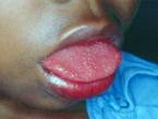
- Erythema, fissures and disappearance of papillary projections of the tongue.
- Traumatic erosions and ulcers, angular cheilitis and lip fissures
- Sleep disturbances due to nocturia, caused by excessive drinking of fluids due to xerostomia
- Chronic esophagitis due to impaired clearance of acids and absence of the regulatory action of saliva
- Weight Loss
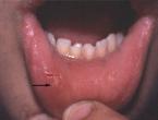
6.2.3.1.3 SALIVARY GLAND ENLARGEMENT
It is observed in 30-50% of patients at some stage in the disease. It is usually hard, diffuse and painless, and evident in the parotid, submandibular and salivary glands. If it is very hard, it can be a sign of malignancy The x-ray with contrast medium injection into the salivary ducts can show sialectasis of the peripheral branches and the MRI, diffuse enlargement of the glands with or without sialectasis.
6.2.3.2 SYSTEMIC MANIFESTATIONS
- Fatigue
- Fever
6.2.3.3 MYOSKELETAL MANIFESTATIONS
Arthralgia with/or without evidence of arthritis(in 50% of patients). Arthropathy is usually symmetrical, intermittent and affects the hands and knees. In primary SS, it is usually nonerosive and nondeforming, despite its association with erosive osteoarthritis. The hand involvement sometimes leads to Jaccoud arthropathy.
Morning stiffness
Myalgia
Muscle weakness
Myositis. It is observed in 2.5-47% of patients with SS. It is usually mild, central and silent and responds to immunosuppressives (Kraus A et al, 1994). Histologically, it is characterized by inflammatory cell infiltrates, even in asymptomatic patients (Bloch KJ et al, 1956).
6.2.3.4 GENITOURINARY MANIFESTATIONS
- Vaginal dryness with dyspareunia (often) (Lehrer S et al, 1994).
- Interstitial cystitis. It is manifested by dysuria, urinary frequency, nocturia and urinary urgency.
- Mild proteinuria
- Renal tubular dysfunction, which can lead to renal tubular acidosis and polyuria due to nephrogenic diabetes insipidus (Pertovaara M et al, 1999)
- Membranoproliferative glomerulonephritis and membranous nephropathy (rare) (Cortez MS et al, 1995)
- Interstitial nephritis
6.2.3.5 MUCOCUTANEOUS DEFECTS
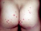
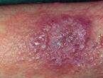
Palpable and non-palpable purpura: more common in patients with positive anti-Ro/SSA and anti-La/SSB, cryoglobulins and/or positive RF. It is apparent in the lower limbs in groups, which may ulcerate. Clinically it is identical to that observed in hypergammaglobulinemic purpura of Waldenstrom.
Urticarial vasculitis, characterized by persistent, probably petechial, painful erythematous papules or plaques.
Digital ulcers.
Rash similar to erythema multiforme.
Nodular vasculitis.
Ring-shaped (doughnut type), erythematous plaques, especially on the face (in the Japanese).
Sweet syndrome lesions (especially on the face) or subacute cutaneous lupus erythematosus.
Raynaud’s phenomenon: It is observed in 13-30% of patients with SS (Kraus A et al, 1992; Garcia-Carassco M et al, 2002). It can be associated with pulmonary fibrosis, arthritis, vasculitis and positive ANA.
Dry nose, vagina and skin. The dry skin may be associated with itching and eczema, especially during the winter months, and skin hyperpigmentation (Ellman P et al, 1951).
6.2.3.6 PULMONARY MANIFESTATIONS
Interstitial lung disease with decreased pulmonary diffusion:It is the most common pulmonary manifestation of SS, but often asymptomatic. The earliest sign is bilateral crepitations.
Pulmonary hyperventilation(Lahdensuo A and Korpela M, 1995)
Increased risk of pulmonary infections(Robinson DA and Meyer CF, 1994).
Upper airway manifestations: Exocrine gland involvement of the upper respiratory tract often leads to dryness of the nasal mucosa, posterior wall of the pharynx, larynx, trachea and bronchi. The most common symptom is a dry cough, which can be persistent and irritating and attributed to asthma or bronchitis.
6.2.3.7 GASTROINTESTINAL MANIFESTATIONS
Dysphagia. It is common in SS. It is usually due to lack of saliva, but also to esophageal dysmotility similar to that observed in SSc (Ramirez-Mata M et al, 1976; Palma R et al, 1994).
Nausea, epigastric pain and dyspepsia(often). The histological examination may show atrophic gastritis with mainly infiltrates of CD4-positive T-lymphocytes.
Achlorhydria and pernicious anemia.
Chronic atrophic gastritis.
6.2.3.8 HEPATIC MANIFESTATIONS
Hepatic disorders.
Primary biliary cirrhosis(Skopouli FN et al, 1994). Patients with primary biliary cirrhosis have an increased incidence of sicca symptoms (Tsianos EV et al, 1990).
Chronic active hepatitis(Skopouli FN et al, 1994).
6.2.3.9 NEUROLOGIC MANIFESTATIONS
Peripheral neuropathy. It is observed in up to 10% of patients with SS. It is manifested by symmetric and sensoryneural polyneuropathy, characterized mainly by sensory disturbances of «glove and socking» distribution (Kumon K et al, 2000).
It presents clinically with anesthesia and dysesthesia of the limbs and is often accompanied by CNS involvement (Alexander EL, 1993). It has a slow and gradual progression and occasionally responds to corticosteroids
Trigeminal Neuralgia: It is the most frequent manifestation of cranial nerve involvement. It usually spares the ophthalmic division, hence the corneal reflex is maintained
Damage to cranial nerve VII(Bell’s palsy)
Damage to cranial nerve VIII, which leads to neurogenic deafness and dysfunction Damage to cranial nerve III, IV or VI, which leads to diplopia.
Autonomic neuropathy.
It is manifested by sympathetic or parasympathetic nervous system dysfunction (pupil and eyelid abnormalities and orthostatic hypotension). It can contribute to abnormal esophageal motility and decreased production of tears and saliva. The nerve biopsy shows axonal degeneration and perivascular inflammatory infiltrates indicative of underlying vasculitis (Mellgren SI et al, 1989).
CNS involvement. It is observed in 20-25% of patients with primary SS (Alexander EL, 1993), or, according to others, less frequently (Binder A et al, 1988; Mellgren SI et al, 1989; Hietaharju A et al, 1990). It is characterized by multifocal recurrent episodes, with long symptom-free intervals , which progressively lead to neurological disorders. Furthermore, it may be manifested by central focal lesions to dementia and conditions similar to multiple sclerosis (Alexander EL, 1993)
CNS INVOLVEMENT MANIFESTATIONS:
- Motor or sensory disturbances (Ohtsuka T et al, 1995), seizures or cerebellar syndromes, as a result of focal lesions
- Encephalopathy, aseptic meningitis or psychiatric disorders ranging from mild impairment of recognition to true dementia (in patients with diffuse brain lesions)
- Transverse or progressive myelopathy due to spinal cord involvement.
- Diplopia, vertigo and ataxia (Gottfried JA et al, 2001)
- Cerebral vasculitis (Berman JL et al, 1990)
- Hypokalemic paralysis associated with renal tubular acidosis (Bartunkova J et al, 1999)
PSYCHIATRIC MANIFESTATIONS:
- Disorders of recognition, with impaired memory and concentration
- Depression and personality disorders (Hietaharju A et al, 1990; Valtysdottir ST et al, 2000)
6.2.3.10 ENDOCRINE DISORDERS
10-70% of patients with primary SS present with structural or hormonal abnormalities of the thyroid or antithyroid antibodies (Bloch KJ et al, 1965; Foster H et al, 1993; Perez B et al, 1995; D 'Arbonneau F et al, 2003 ). According to others, primary SS is not associated with thyroid conditions (Ramos-Casals M et al, 2000).
6.2.4 LABORATORY FINDINGS
- Mild anemia - leukopenia (in around 1/3 of patients)
- Increased ESR (in 80-90% of patients). CRP is usually within normal limits
- Hyperamylasemia
- Polyclonal hypergammaglobulinemia (IgG) (in 80% of patients)
- Positive ANA and RF
- Positive anti-Ro (SS-A) (40-45%) and anti-La (SS-B) (50%), at high titers with a spotted or nuclear immunofluorescence pattern. Anti-Ro antibodies are associated with anemia, leukopenia, lymphopenia, cryoglobulinemia and vasculitis.
- Positive lupus cells (in 15% of patients, even without SLE)
- Positive antibodies to thyroglobulin, mitochondria, smooth muscle fibres and salivary ducts (in 2/3 of patients).
6.2.5 IMAGING MODALITIES
Sialography: It can show sialectasis.
Sialometry: It identifies the total amount of produced saliva or individialized secretions of the salivary glands at rest or after stimulation and the effectiveness of treatment with sialagogue agents. The saliva should be collected for at least 5 min and often up to 15 min. Quantity reduction to <0.1 mL/min at rest is indicative of hypofunctioning salivary glands, while the total amount of produced saliva after stimulation amounts at <0.5 mL / min.
Sialochemistry: It often shows increased secretory IgA, lactoferrin, total proteins and sodium and chlorine ions, as well as decreased lysozyme and potassium and phosphate ions.
Schirmer test: It serves to assess the production of tears by the lacrimal glands. A thin strip of paper is placed beneath the lower eyelid and after 5 min the wetted length is measured. If it is <5 mm, it is an indication of decreased tear production. The salivary gland function can be roughly estimated with the following questions:
- Can you swallow dry foods, eg crispbread, without drinking water?
- Do you feel your mouth is dry when you eat your food?
- Do you have difficulty swallowing some foods?
- Do you think your mouth is dry and does not have much saliva?
Positive answers to all these above questions suggest hypofunction of the major salivary glands.
Scintigraphy with technetium Tc 99: It shows delayed retention of the radiopharmaceutical associated with histopathological changes.
Magnetic resonance imaging: It reveals the glandular parenchyma, particularly the cystic or solid masses, and determines the volume of the glands.
6.2.6 HISTOLOGICAL FINDINGS
Kidneys: Interstitial and peritubular infiltrates of lymphocytes, histiocytes and plasma cells.
Salivary glands:
- Lymphocytic infiltration of glandular acini of the lacrimal and/or salivary glands, mainly by CD4 + T-lymphocytes, as well as by activated B-lymphocytes and, less often, plasma cells. These infiltrates are focal and surrounded by normal acini. The discovery of more than one foci or ≥ 50 inflammatory cells in glandular tissue area of 4 mm2 is indicative of SS. The greater the number of foci, the higher the association with the disease.
- Peritubular and perivascular hyaline depositions
- Tubular epithelium hyperplasia and myoepithelial cells concentrated in epimyoepithelial islets in the lymphoid tissue. Unlike the parotid lesions, epimyoepithelial islets are rarely observed in the lymphocyte surface.
- Formation of germinal centers.
The histologic findings of salivary gland biopsy are very useful for the diagnosis of SS. The salivary gland biopsy is preferred to that of parotids, which can damage the facial nerve. The biopsied sample should include 5-10 acini of minor salivary glands. If there is parotid enlargement, a fine-needle biopsy or open biopsy may be needed to rule out the probability of cystic or neoplastic disease. In some cases, the estimation of the clonality of lymphocytic infiltrates by immunohistochemical studies or studies of genetic upgrading may be necessary to exclude low-grade B-cell lymphoma. The major gland enlargement may be associated with benign lymphoepithelial lesions. The lesions are characterized by dense lymphocytic infiltrations associated with acinar salivary gland destruction while the tubular epithelium is maintained.
6.2.7 DIAGNOSIS
The probability of SS should be considered in children who have:
- Foreign body feeling in the eye
- Chronic xerostomia and xerophthalmia
- Recurrent parotid gland enlargement (with/or without sicca complex)
- Early and advanced dental caries associated with xerostomia
- Increased ESR, hypergammaglobulinemia or positive RA test, positive ANA and anti-Ro (SSA) or anti-La (SSB) antibodies.
The diagnosis of Sjogren’s syndrome in children may be based on the proposed criteria of Deprettere et al. (TABLE 73) (Deprettere AJ et al, 1988). These criteria are mainly used in adults, but can also be used in children.
TABLE 73
PROPOSED DIAGNOSTIC CRITERIA FOR JUVENILE SJOGREN’S SYNDROME (Deprettere AJ et al, 1988)
- Keratoconjunctivitis confirmed by Schirmer test and the quantitative test of Rose Bengal staining.
- Xerostomia confirmed by reduced basal and post-stimulatory salivation.
- Lymphocytic infiltration of a minor salivary gland sample containing at least 2 foci / 4 mm2.
- Laboratory indication of a systemic autoimmune disease based on RF in titre of ≥ 1/160, positive ANA in titre of ≥ 1/160 or positive anti-ENA
6.2.8 DIFFERENTIAL DIAGNOSIS
Infections
- Herpes simplex
- Infectious parotitis (mumps virus, streptococcus, staphylococcus, HIV-1, Epstein-Barr, coxsackie)
- AIDS
- Mononucleosis
- Epstein-Barr
Rheumatic diseases
- Mixed connective tissue disease
- Systemic lupus erythematosus
- Systemic scleroderma
Oral diseases
- Gingivitis/periodontitis
- Oropharyngeal candidiasis
- Recurent bacterial sialadenitis
- Superficial mucocele
- Difficulty in fitting dental prostheses
- Other
- Dryness of nasal and vaginal mucosa and skin
- Use of anticholinergics and antidepressants
- Vasculitis and thrombophlebitis
- Recurrent (non-autoimmune) juvenile parotitis
- Tumors
- Sarcoidosis
- Melkersson-Rosenthal syndrome
6.2.9 COMPLICATIONS
- Renal-pancreatic calcifications (Grindulis KA and Roy S, 1987)
- Chronic thyroiditis (Kobayashi I et al, 1996)
- Interstitial nephritis
- Inflammation of sweat glands (Kobayashi I et al, 1996)
6.2.10 TREATMENT
6.2.10.1 CHOLINERGIC AGONISTS
INDICATIONS: Stimulation of saliva production
Pilocarpine. The dose has not been determined. Titration of doses from 2.5 mg per os bid.
Cevimeline. Dose (in adults) 30 mg per os tid. In children it has not been determined.
6.2.10.2 NON-STEROIDAL ANTI-INFLAMMATORY DRUGS
INDICATIONS: Articular manifestations
Naproxen:
- Children <2 years: Not determined
- Children> 2 years: 10-20 mg/kg/24h per os, in divided doses bid / tid; The dose should not exceed that of adults
6.2.10.3 STEROIDS
Corticosteroids per os
INDICATIONS: Polyarthritis uncontrolled by NSAIDs, extraglandular manifestations (interstitial pneumonitis, glomerulonephritis, vasculitis, pseudolymphoma, malignancy).
DOSE: Prednisone 1-2 mg/kg/24h per os.
Intravenous infusions of methylprednisolone
INDICATIONS: Renal involvement (Rosenberg AM et al, 1990)-CNS (Gottfried JA et al, 2001).
DOSE: 30 mg/kg/24h x 3 consecutive days.
6.2.10.4 ANTIMALARIAL
Hydroxychloroquine: It improves significantly dry eyes and xerostomia and systemic manifestations (arthralgias, myalgias and lymphadenopathy) of the disease after 1-3 years of treatment (Fox RI et al, 1996). In combination with small doses of corticosteroids, it mainly improves the extraglandular manifestations and much less xerostomia and dry eyes in children with primary Sjogren’s syndrome (Ostuni PA et al, 1996). It can help to reduce the dose of corticosteroids in patients with visceral involvement (vasculitis, skin lesions, pneumonitis, neuropathy, neuritis). According to others, it does not significantly improve the glandular and extraglandular manifestations of the disease (Kruize AA et al, 1993; Tishler JM et al, 1999).
Chloroquine - IFN-a2: In patients with Sjogren’s syndrome, IFN-alpha2 improves the function of the lacrimal and salivary glands in 67% and 61%, compared with 15% and 18% with chloroquine, respectively (Ferraccioli GF et al, 1996).
RECOMMENDATIONS: Hydroxychloroquine may improve the extraglandular manifestations (arthritis, arthralgia, myalgia, lymphadenopathy) of primary Sjogren’s syndrome, but it is debatable whether it improves xerostomia and xerophthalmia, hence the potential benefit of its use should be weighed compared with the risks (especially retinopathy) of long-term treatment with antimalarials.
6.2.10.5 IMMUNOSUPPRESSIVE AGENTS
Methotrexate: It reduces symptoms, but not the objective parameters, of sicca syndrome (Skopouli FN et al, 1996) and is effective in arthritis. It can be combined with other DMARDS (hydroxychloroquine, sulfasalazine, penicillamine).
Cyclosporine: It improves xerostomia subjectively, but not objectively (Drosos AA et al, 1986).
Biological agents (infliximab - etanercept): They do not seem to be effective in primary SS (Mariette X et al, 2003; Zandbelt MM et al, 2004).
6.2.10.6 PREVENTION - TREATMENT OF SJOGREN’S SYNDROME MANIFESTATIONS
Xerostomia
- Sielagogues eg pilocarpine (Salagen) 5 mg 3-4 times daily or cevimeline 30 or 60 mg 3 times daily
- Chewing mastic gum, maltose lozenges
- Frequent intake of water and other liquids
- IFN-a, administered orally, is debatable whether it increases saliva production
- Keratoconjunctivitis sicca
- Artificial tears with hypromellose, methylcellulose, or androgens
- Acetylcysteine for mucus filaments accumulated in the eyes
- Cyclosporine 0.05%
- Corticosteroids without preservatives and calcium ingredients
Dental caries - gingivitis - periodontitis
- Brushing the teeth 2 times a day with fluoride toothpaste and daily cleaning with dental floss
- Cleaning the teeth 3-4 times a year
- Oral rinses with chlorhexidine gluconate for 2 weeks if there is a large number of Streptococcus mutans in saliva (> 1 X 10⁶/mL saliva).
- Reduced consumption of refined sugar, simple carbohydrates and acidic foods and drinks
Oropharyngeal fungal infections
- Local (suspensions or sucrose-free lozenges) and systemic antifungal agents.
- Daily cleaning of artificial dentures with chlorhexidine 0.2% solution
- Good hygiene and frequent wetting and lubrication of the mouth
Treatment of oral candidiasis
- Chlortrimazole topically and orally for 1-2 weeks (for angular cheilitis)
Arthralgias/myalgias: Simple analgesics and/or NSAIDs. Hydroxychloroquine, if response is inadequate.
Cutaneous lesions
- Antimalarials (for maculopapular rash)
- Local corticosteroids, except for the face, where topical tacrolimus is recommended to prevent thinning of the skin caused by corticosteroids
Serious cardiopulmonary manifestations: Corticosteroids with/or without immunosuppressive agents per os (chlorambucil or azathioprine) or IV cyclophosphamide infusions, at a dose of 500-1.000 mg/month X 6 months.
Glomerulonephritis: Treatment is the same as in renal SLE.
Gastrointestinal manifestations: Proton pump inhibitors, prokinetic agents.
Mononeuritis multiplex: Corticosteroids and immunosuppressants (such as in the treatment of polyarteritis).
Sensory neuropathes in the hypergammaglobulinemia purpura:
- Treatment of underlying condition (hyperlipidaemia, endocrine disorders, hypertension)
- Gabapentine.
Peripheral motor neuropathies: IVIG and/or immunosuppressants (Takahashi Y et al, 2003; Levy Y et al, 2003)
Autonomic neuropathy: Fluorocortisone acetate or midodrine.
CNS manifestations: Corticosteroids, cyclophosphamide and other immunosuppressants.
6.2.10.7 RECOMMENDATIONS IN PATIENTS WITH SJOGREN’S SYNDROME
- Drink plenty of fluids with meals to improve chewing, taste and swallowing of foods.
- Eat dairy products daily, mostly low fat milk, yoghurt and cheese. Milk lubricates the mouth more and the cheese has beneficial effects against dental caries.
- Eat foods at moderate temperatures (not too cold - not too hot)
- If you swallow with difficulty, you can liquify or grind the food
- If you need to increase calorie intake due to nutritional disease, you can take liquid dietary supplements
- Limit the consumption of sugary foods and soft drinks between meals.
- Alternatively, you can use sweeteners, if tolerated, such as aspartame, saccharin, sorbitol and xylitol.
WHAT PATIENTS WITH SJOGREN’S SYNDROME SHOULD AVOID
- Dry, crunchy foods, because they are swallowed with difficulty and irritate the gastrointestinal mucosa
- Spicy or acidic foods and drinks
- Simple carbohydrates such as sucrose and well-refined processed foods, eg cakes and biscuits, to reduce the risk of developing caries.
- Beware of artificial sweeteners because they may cause abdominal discomfort
- Alcoholic and caffeinated (coffee, tea, cola) beverages because they aggravate xerostomia.
- Oral washes with fluids containing alcohol, because they dry the oral mucosa
- Smoking
- Exposure to windy and dry environment with low humidity
- Saliva substitutes (eg carboxymethylcellulose), because they usually are not effective.
6.2.11 PROGNOSIS
Primary SS usually has a good prognosis, unless it is accompanied by severe extragrandular manifestations. The prognosis of secondary SS depends on the primary disease. Patients with SS, especially primary SS, have an increased risk of developing lymphoma (Ramos-Kasals M etg al, 2002), although the overall duration of survival is similar to subjects without SS (Ioannidis JP et al, 2002). The extragrandular lymphoid hyperplasia (ie lymphadenopathy and hepatosplenomegaly), observed in SS, sometimes mimics pseudolymphoma, but may also be a precursor of malignant disease (non-Hodgkin lymphoma, Waldenstrom’s macroglobulinemia, B-cell lymphoma).
ΑΠΟΔΟΣΗ ΣΤΑ ΑΓΓΛΙΚΑ Ναταλί Μαίση, Ιατρός

Θέματα
Συλλογή Διαφανειών
 JPEG.jpg)
 JPEG.jpg)
 Πόδια JPEG.jpg)
 Γόνατα JPEG.jpg)
 Χέρια JPEG.jpg)
Συλλογή Φωτογραφιών

 Image Collection.jpg)

.jpg)
.jpg)
.jpg)
 KONSTANTINA Πρόσωπο (1).jpg)

.jpg)
.jpg)
.jpg)
 Φωτο + αα.jpg)


.jpg)

 JPEG.jpg)
 Αγκώνας JPEG.jpg)
 Χέρια JPEG.jpg)
_0.jpg)
_0.jpg)
 Μαλτέζος Πρόσωπο (4).jpg)
 KONSTANTINA Χέρια (3).jpg)
.jpg)
.jpg)
 JPEG.jpg)
 JPEG.jpg)
 JPEG.jpg)
 JPEG.jpg)
 JPEG.jpg)
 JPEG.jpg)
 JPEG.jpg)
 JPEG Γόνατα.jpg)
 JPEG Αγκώνες (2).jpg)
.jpg)
 Σημείο αντίχειρα.jpg)
 άγνωστης αιτιολογίας.jpg)
 Φωτο (2) Παθητική ανάταξη (1).jpg)

.jpg)

.jpg)
.jpg)
.jpg)

.jpg)
.jpg)
 Χέρια Φωτο.jpg)
 JPEG.jpg)
 Χέρια Φωτο.jpg)
.jpg)
.jpg)
 Σημείο Gottron (1).jpg)
 Σημείο Gottron (2).jpg)
 Υποδόριες ασβεστώσεις (1).jpg)
 Υποδόριες ασβεστώσεις (2).jpg)
 Χέρια JPEG.jpg)
 Χέρια JPEG.jpg)
 Χέρια JPEG.jpg)
 Χέρια JPEG.jpg)
 Χέρια JPEG.jpg)
 Χέρια JPEG.jpg)
 Χέρια JPEG.jpg)
 Χέρια JPEG.jpg)
 Χέρια JPEG.jpg)
 Χέρια JPEG.jpg)
 Χέρια JPEG.jpg)
 Χέρια JPEG.jpg)
 Εξάνθημα JPEG.jpg)
 JPG.jpg)
 JPG.jpg)
 JPG_0.jpg)
 JPG_0.jpg)
 JPG.jpg)
 Οσφύς JPEG.jpg)
 Οσφύς JPEG.jpg)
 Φωτο.jpg)
 Φωτο.jpg)
 Βραχυδακτυλία JPEG.jpg)


_2.jpg)
_0.jpg)
 Αρθρίτιδα χεριών.jpg)
 Δερματικό εξάνθημα.jpg)
.jpg)
_0.jpg)
.jpg)
 Υποδόριες ασβεστώσεις (1)_0.jpg)
 Υποδόριες ασβεστώσεις (2)_0.jpg)
_0.jpg)
 Χέρια Φωτο_0.jpg)
 Χέρια Φωτο_0.jpg)
 Χέρια JPEG.jpg)
 Aρθροπάθεια.jpg)
 Αγγειοκερατώματα.jpg)
_1.jpg)
.jpg)
.jpg)
_2.jpg)
 (2).jpg)
_1.jpg)
.jpg)


Βότανα-Φυσικές ουσίες
